ADVANCE TESTING
RETINAL IMAGING
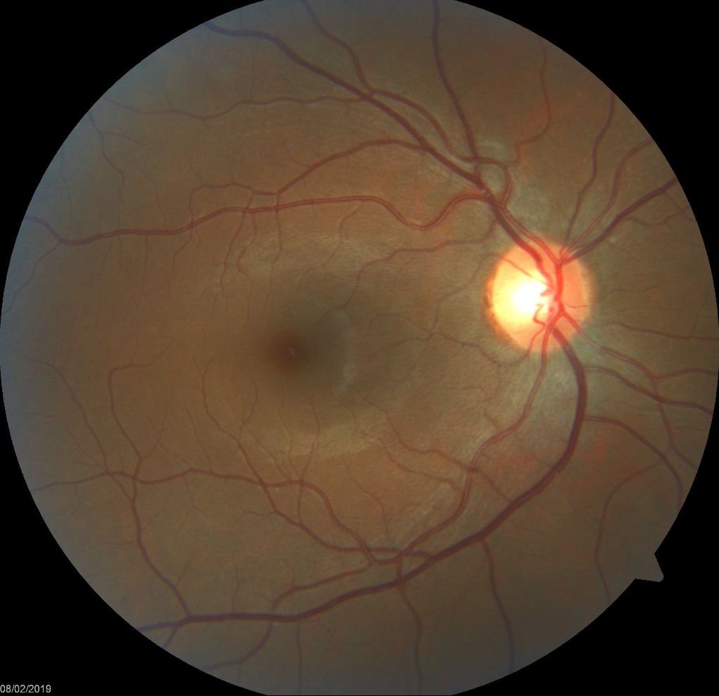
Retinal Imaging is a picture taken of the back of the eye, specifically the retina, optic nerve, macula and the blood vessel. Phots helps us monitor for changes over time. Certain conditions such as Glaucoma, Macular Degeneration, Diabetic Retinopathyand even Myopia can show slow progressive changes to the retina tissue over time and imaging makes it easier to track these changes.
Dr. Patel recommends Retinal Imaging to all age groups especially in patients with risk factors and family history to certain eye conditions.
OCT – OPTICAL COHERENCE TOMOGRAPHY
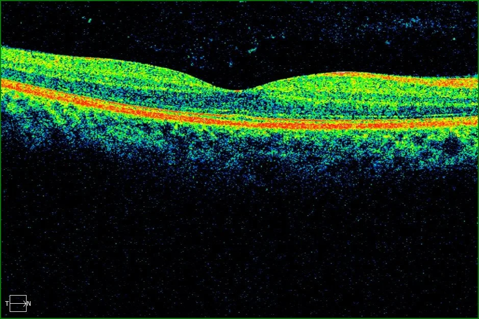
OCT is like a an ultrasound ( but instead of using soundwaves, it uses light waves) to illuminate and scan your retina. It is non-invasive imaging of the difference layers of the back of the eye (retina) to help detect any damages of changes due to conditions such as glaucoma, diabetic retinopathy, macular degeneration and so much more.
VISUAL FIELD
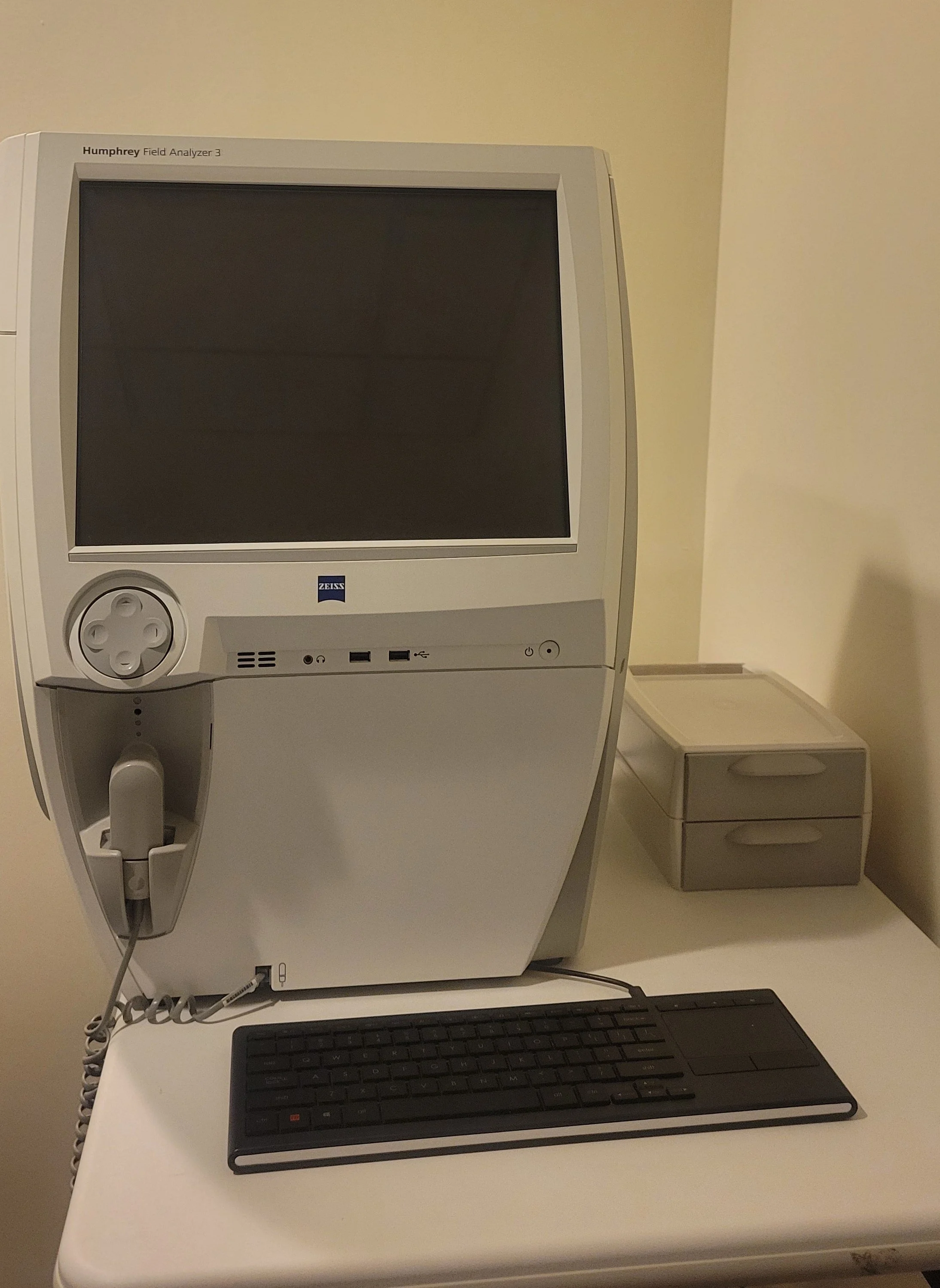
VISUAL FIEVISUAL FIELD TEST – This test maps both your central and peripheral vision, giving insight into how you see the world. It's a vital tool for detecting conditions such as glaucoma, strokes, brain tumors, and other neurological issues.
The visual field test helps identify any defects or abnormalities along the visual pathway — starting from the front (the optic nerve, which transmits visual information from the eyes to the brain) to the back (the occipital lobe, where that information is processed).
In addition to medical use, this test may also be required for certain Vision Screening, such as policing or pilot, or by regulatory bodies like the Ministry of Transportation to ensure adequate vision for driving or job performance.
CORNEAL TOPOGRAPHY
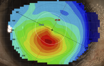
Corneal Topography is a quick, non-invasive test that measures and creates a detailed map of the shape and curvature of your cornea—the clear front surface of the eye where contact lenses rest.
The cornea plays an important role in vision by helping to focus light as it enters the eye. Because of this, even small changes in the cornea's shape can affect how well you see.
We use corneal topography to help detect and monitor certain eye conditions, such as Keratoconus, and to assist with fitting specialty contact lenses, including Orthokeratology lenses. It is also commonly used by eye specialists when assessing patients for laser vision correction procedures and other corneal treatments.
The test is painless, does not involve touching the eye, and provides valuable information about the health and shape of your cornea.